
Primary mouse microglia cells play a critical role in the central nervous system (CNS), acting as the resident immune cells of the brain. These cells are instrumental in maintaining CNS homeostasis, responding to injury, and modulating inflammatory responses. Microglia cells, which originate from yolk-sac progenitors during early development, persist throughout life and constantly survey their environment for potential threats, including infections and tissue damage. Their relevance to neurodegenerative diseases, such as Alzheimer’s and Parkinson’s, has made them a focus of extensive scientific investigation. This article explores the significance of primary mouse microglia cells in research, emphasizing their importance in both basic neuroscience and translational studies.
Microglial Functions in the CNS
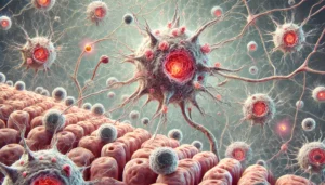
Microglia are dynamic cells that alter their morphology and function in response to environmental cues. In the healthy brain, microglia remain in a “resting” state, constantly scanning their surroundings through highly motile processes. When injury occurs, they rapidly shift to an activated state, migrating to the injury site to clear debris and release pro-inflammatory cytokines (NIH) (NINDS). This unique adaptability makes microglia a subject of interest in studies of neuroinflammation (NIA).
Isolation and Cultivation of Primary Mouse Microglia Cells
The isolation of primary microglia from mouse brains has become a standard method for studying their function in vitro (NCBI). Techniques typically involve dissociating brain tissue and separating microglia using density gradient centrifugation (CDC). Specialized media are used to promote their growth while preserving their distinct phenotypic characteristics (NIH).
This isolation process provides researchers with pure microglial populations, allowing for detailed analysis of their behavior under experimental conditions (PubMed). Studies using primary mouse microglia have yielded insights into roles in synaptic pruning, phagocytosis, and the modulation of neuronal activity (NIH) (NCBI).
Microglia and Neurodegenerative Diseases
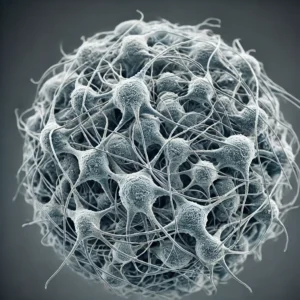
Microglia are key players in neurodegenerative diseases, where they exhibit chronic activation. In conditions like Alzheimer’s disease, they are implicated in amyloid plaque formation and can exacerbate neuroinflammation (Alzheimer’s Association) (NIA). Similarly, in Parkinson’s disease, microglial activation contributes to dopaminergic neuron loss (NINDS). Their involvement in neurodegenerative processes makes them a promising target for therapeutic interventions (NIH).
Genetic Tools and Disease Modeling
Primary mouse microglia cells are frequently used in conjunction with transgenic mouse models to study the genetic underpinnings of neurodegenerative disorders (NCBI). For example, knockout models help researchers understand how specific genes influence microglial function (EMBL). These models are critical for dissecting molecular pathways involved in neurodegeneration and testing potential therapies (NINDS).
Additionally, CRISPR-Cas9 gene-editing technologies have allowed researchers to modify microglia cells, further expanding the possibilities for disease modeling (NIH) (CDC).
Inflammatory Responses and Microglial Activation
Understanding how microglia respond to inflammatory stimuli is crucial for developing treatments for neuroinflammatory disorders like multiple sclerosis (MS). In MS, activated microglia contribute to demyelination and axonal damage (NIH) (PubMed). Studies using primary mouse microglia cells have been pivotal in elucidating the pathways involved in inflammation and identifying anti-inflammatory therapy targets (CDC) (NINDS).
Microglia and the Blood-Brain Barrier
Research has also focused on the interaction between microglia and the blood-brain barrier (BBB). Microglia help maintain BBB integrity and mediate immune responses during CNS infections (NINDS). Disruption of this interaction can lead to increased BBB permeability, allowing harmful substances to enter the brain (NCBI).
Applications in Drug Screening and Toxicology
Primary mouse microglia cells are valuable in drug screening and toxicology studies. Their ability to secrete cytokines, chemokines, and other signaling molecules upon activation makes them useful for testing anti-inflammatory drugs and neuroprotective agents (CDC) (NCBI). Moreover, microglia are used to assess the neurotoxic effects of environmental toxins and pharmaceuticals (NIH).
Conclusion
Primary mouse microglia cells are indispensable for advancing our understanding of neuroimmunology. Their involvement in various CNS processes and disease pathology makes them powerful tools for brain research and the development of new therapies. As the research landscape evolves, these cells will remain central to unraveling the complexities of the brain’s immune system.
For further information, you can visit trusted resources like NIH, NINDS, CDC, NCBI, Alzheimer’s Association, and PubMed. These sources offer comprehensive insights into the applications of primary microglia cells in neuroscience and neuroimmunological research.